

2026-11-18
模型相关
近期,美国华盛顿大学医学院(Washington University School of Medicine)Marco Colonna团队联合David M. Holtzman团队,在《Science》上发表题为"Targeting amyloid-β pathology by chimeric antigen receptor astrocyte (CAR-A) therapy"的研究论文。该研究将CAR技术应用于星形胶质细胞,构建了靶向 Aβ 的嵌合抗原受体星形胶质细胞(CAR-A),借助可穿透血脑屏障的 AAV 载体,通过单次静脉注射即可实现脑内星形胶质细胞的持续性基因改造。在阿尔茨海默病小鼠模型中,CAR‑A 能够显著降低 Aβ 斑块负荷、改善斑块相关神经病理,可长期发挥 Aβ 清除作用,为阿尔茨海默病的疾病修饰治疗开辟全新思路。
研究背景:现有 AD 抗体疗法仍存诸多瓶颈
阿尔茨海默病(AD)是造成痴呆的首要病因,核心病理特征为脑内 Aβ 淀粉样斑块沉积,继而诱发 tau 蛋白介导的神经退行性病变。目前已经获批的 Aβ 单抗药物(Lecanemab、Donanemab)可依靠小胶质细胞的抗体依赖性细胞吞噬(ADCP)通路清除 Aβ 斑块,实现临床疾病进展延缓,但该类疗法仍存在难以忽视的短板:
血脑屏障穿透难题:需要超高给药剂量,长期治疗成本高昂;
安全性风险:容易诱发淀粉样蛋白相关影像学异常(ARIA),需要频繁监测患者;
治疗窗口受限:晚期干预对认知改善有限,高度依赖最佳干预时机;
机制局限:治疗完全依赖 FcRγ 信号,难以灵活调控细胞应答;
基于上述痛点,研究团队提出全新设想:不依靠反复注射外源抗体,而是对大脑内源细胞进行基因工程改造,让脑细胞自身获得清除 Aβ 斑块的能力。
核心发现:改造星形胶质细胞,打造脑内 “清斑能手”
星形胶质细胞是脑内数量丰富且具备吞噬功能的胶质细胞,本研究将其作为改造靶点,构建抗 Aβ CAR‑A;利用 AAV‑PHP.eB 载体实现非侵入式全脑星形胶质细胞靶向表达,达成单次给药、脑内长期清除 Aβ的治疗效果。
关键设计一:体外多构型筛选,锁定最优 CAR‑A 分子
研究构建 4 种携带不同胞内信号域(Megf10、Dectin1、MERTK、CD3ζ)的 CAR 骨架,分别搭配 Crenezumab、Aducanumab 两种抗 Aβ 单链可变片段(scFv)。体外细胞实验证实,CAR‑A 能够特异性识别、吞噬并降解 Aβ 寡聚体,不会非特异性摄取 tau、α‑突触核蛋白、髓鞘碎片等其他蛋白聚集体。经过功能筛选,确定Crene‑Megf10、Adu‑Dectin1两个最优构型开展后续体内动物实验。
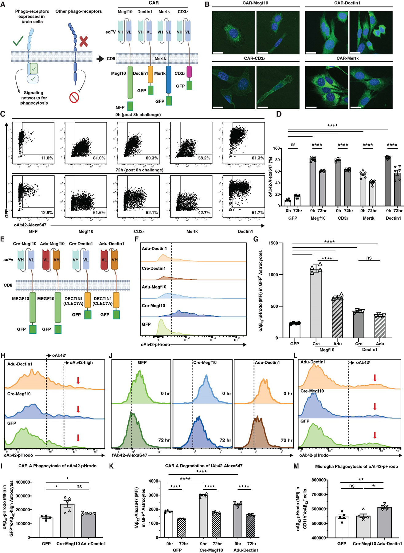
图1. CAR-A的设计原理及体外功能验证(A) Schematic representation of the rationale used to select appropriate phagocytic receptors (left) and the designs of CARs with different intracellular signaling domains (right). (B) Confocal imaging of immortalized astrocytes transduced with the Crene-CARs, nuclei are stained with DAPI (blue). Scale bar: 20 μm. (C) Flow cytometry plots of oligomeric Aβ42–647 (oAβ42–647) uptake by Lonza primary astrocytes at 8 hours phagocytosis and 72 hours post phagocytosis. (D) Quantifications of the percentage of oAβ42–647 CAR astrocytes at 0h and 72h post 8 hours of phagocytosis. N=6. (E) Schematic representation of CAR-Megf10 and CAR-Dectin1 with different scFvs. (F) Representative flow cytometry histograms of oAβ42-pHrodo uptake by Crene-Megf10, Adu-Megf10, Crene-Dectin1 and Adu-Dectin1 astrocytes at 8 hours. (G) The mean fluorescence intensity (MFI) of oAβ42-pHrodo uptake by Crene-Megf10, Adu-Megf10, Crene-Dectin1 and Adu-Dectin1 astrocytes at 8 hours. N=4. (H) Representative flow cytometry histograms of oAβ42-pHrodo uptake by GFP-, Crene-Megf10-, and Adu-Dectin1-expressing astrocytes in the mixed glial culture at 8 hours. Two populations of oAβ42+ astrocytes are indicated by labels in the panel. (I) Quantifications of the MFI of Aβ42-pHrodo uptake by GFP−, Crene-Megf10−, and Adu-Dectin1-expressing astrocytes in the mixed glial culture at 8 hours. (J) Representative flow cytometry histograms of fibrillar Aβ42–647+ uptake by GFP−, Crene-Megf10−, and Adu-Dectin1-expressing astrocytes at 0h and 24h post 8 hours of phagocytosis. (K) Quantifications of the MFI of fibrillar Aβ42–647+ uptake by GFP−, Crene-Megf10-, and Adu-Dectin1-expressing astrocytes at 0h and 24h post 8 hours of phagocytosis. (L) Representative flow cytometry histograms of oAβ42-pHrodo uptake by microglia in each mixed glial culture at 8 hours. Populations of oAβ42+ microglia are indicated by labels in the panel. (M) Quantifications of the MFI of Aβ42-pHrodo uptake by microglia in the mixed glial culture at 8 hours. N=5. All data presented as mean ± SEM with technical replicates except that Fig. 1I, K, M with biological replicates.(图源:Science. 2026 March 05; 391(6789): eads3972. doi:10.1126/science.ads3972.)
关键设计二:AAV 非侵入递送,实现星形胶质细胞特异性表达
研究团队将Cren-Megf10和Adu-Dectin1两种CAR构建体装入AAV-PHP.eB载体(一种可穿越血脑屏障的AAV血清型),由GFAP启动子驱动,实现星形胶质细胞特异性表达。通过静脉注射即可实现全脑递送,无需颅内注射。
核心成果一:晚期治疗——单次给药减少约50%淀粉样负荷
选用 6 月龄已形成大量 Aβ 斑块的 5xFAD 小鼠,进行单次静脉给药,3 个月后评估病理改变。结果显示,相较于对照组,Crene‑Megf10 与 Adu‑Dectin1 两种 CAR‑A 均使小鼠全脑淀粉样负荷下降约 50%,皮层、海马脑区斑块负荷显著降低,ELISA 检测也证实脑内致病性不溶性 Aβ42 聚集体水平明显下降。
神经病理检测表明,两种 CAR‑A 均可减少斑块周边营养不良神经突起;其中 Crene‑Megf10 还可以部分改善突触后标志物 PSD95 的表达水平。
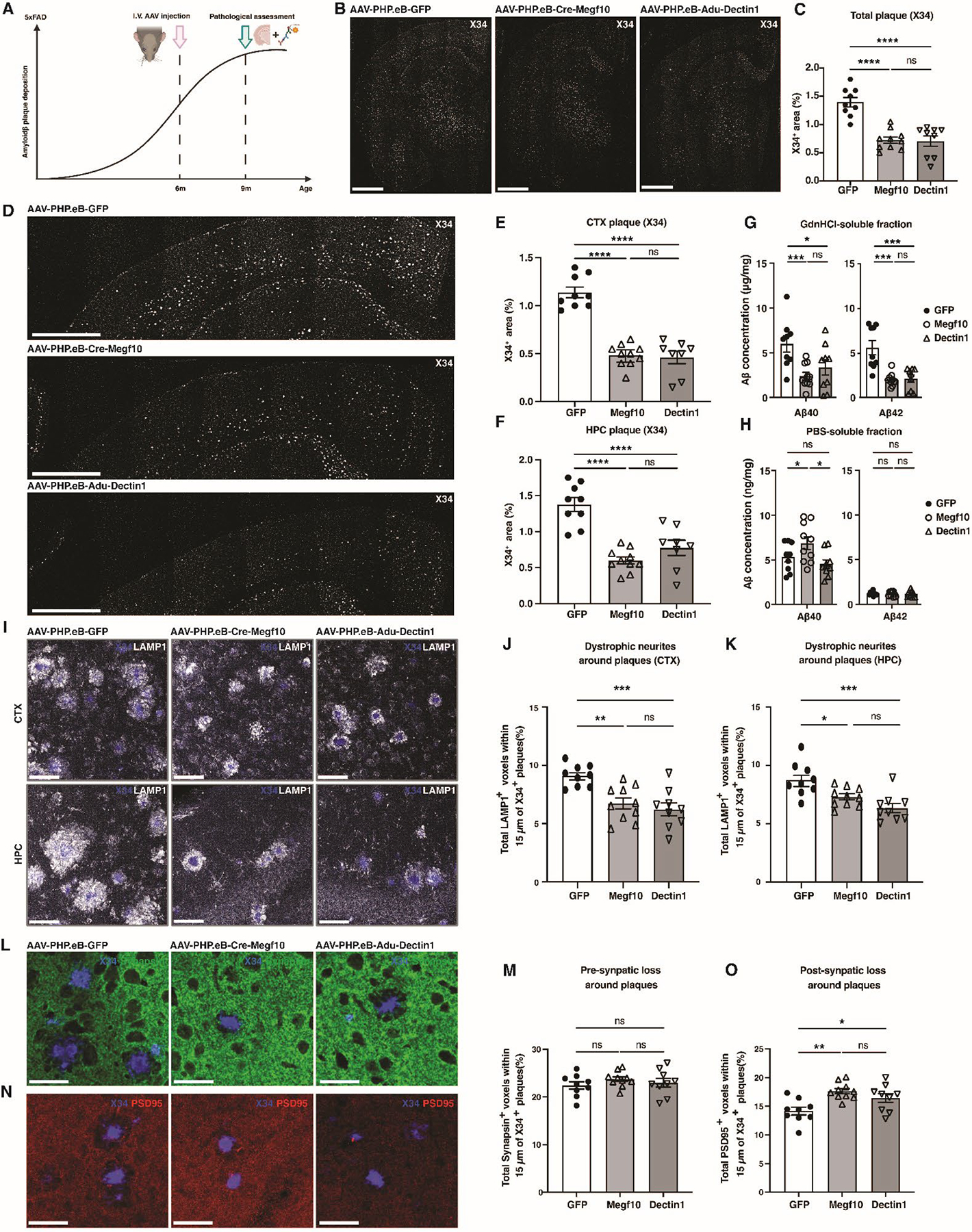
图2. 晚期CAR‑A治疗减少Aβ病理(A) Experimental timeline for AAV injection and pathological assessment in 5xFAD mice post plaque formation. (B) Representative X34 staining of amyloid plaques in coronal brain sections. Scale bars: 1000 μm. (C) Quantification of total X34+ plaque area in coronal sections. (D) Representative X34 staining of amyloid plaques in cortical sections from mice at 9 months of age, injected with AAV-PHP.eB -GFP, AAV-PHP.eB-Crene-Megf10, or AAV-PHP.eB-Adu-Dectin1. Scale bars: 1000 μm. (E and F) Quantification of X34+ plaque area in cortex (E) and hippocampus (F). (G and H) ELISA results of Aβ40/42 in the GdnHCl soluble (G) or PBS soluble (H) fractions from cortical tissues of treated 5xFAD mice at 9 months of age. (I) Representative confocal images of X34+ (blue) plaques and LAMP1 (white) staining in cortex and hippocampus. Scale bars: 30 μm. (J and K) Quantification of total LAMP1+ voxels within 15μm around X34+ plaques (%) in the cortex (J) or hippocampus (K) of treated 5xFAD mice at 9 months of age. (L) Representative images and (M) quantification of total Synapsin+ voxels (green) within 15μm around X34+ plaques (%) in the cortex of treated 5xFAD mice at 9 months of age. (N) Representative images and (O) quantification of total PSD95+ voxels (red) within 15μm around X34+ plaques (%) in the cortex of treated 5xFAD mice at 9 months of age. Scale bars: 30 μm. Data in D, E, G, H, J, K, M, O are presented as mean ± SEM. N=9 for GFP, N=10 for Megf10, and N=9 for Dectin1. Statistical significance was calculated using one-way ANOVA with either Dunnett correction or Tukey correction for multiple comparison: *P < 0.05, **P < 0.01, ***P < 0.001, ****P < 0.0001, ns: not significant.(图源:Science. 2026 March 05; 391(6789): eads3972. doi:10.1126/science.ads3972.)
核心成果二:早期干预——近乎阻断斑块形成
在尚未出现 Aβ 斑块的 2.5 月龄 5xFAD 小鼠开展预防性给药,待小鼠成长至 5 月龄开展病理评估。实验证实两种 CAR‑A 均可抑制早期 Aβ 斑块生成,Crene‑Megf10 组脑内几乎观察不到致密淀粉样斑块;脑内致病性不溶性 Aβ42 水平显著下降,Crene‑Megf10 组中该指标低于检测下限。同时,两种 CAR‑A 均可减轻残存斑块周围神经突起损伤,改善 Synapsin、PSD95 突触标志物,起到突触保护作用。
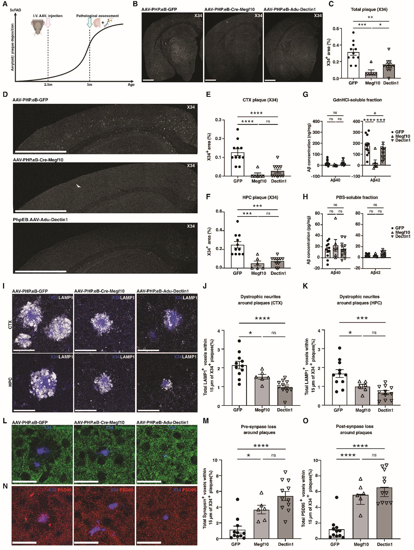
图3. 早期CAR‑A干预预防斑块形成(A) Experimental timeline for AAV injection and pathological assessment in 5xFAD mice prior to plaque formation. (B) Representative X34 staining of amyloid plaques in coronal brain sections. Scale bars: 1000 μm. (C) Quantification of total X34+ plaque area (%) in coronal sections. (D) Representative X34 staining of amyloid plaques in cortical sections from mice injected with AAV-PHP.eB -GFP, AAV-PHP.eB -Crene-Megf10, or AAV-PHP.eB-Adu-Dectin1 at 5 months of age. Scale bars: 1000 μm. (E and F) Quantification of X34+ plaque area in cortex (E) and hippocampus (F). (G and H) ELISA results of Aβ40/42 in the GdnHCl soluble (G) or PBS soluble (H) fractions from cortical tissues of treated 5xFAD mice at 5 months of age. (I) Representative confocal images of X34+ (blue) plaques and LAMP1 (white) staining in cortex and hippocampus. Scale bars: 30 μm. (J and K) Quantification of total LAMP1+ voxels within 15μm around X34+ plaques (%) in the cortex (J) or hippocampus (K) of treated 5xFAD mice at 5 months of age. (L) Representative images and (M) quantification of total Synapsin+ voxels (green) within 15μm around X34+ plaques (%) in the cortex of treated 5xFAD mice at 5 months of age. (N) Representative images and (O) quantification of total PSD95+ voxels (red) within 15 μm around X34+ plaques (%) in the cortex of treated 5xFAD mice at 5 months of age. Scale bars: 30 μm. Data in C, E-H, J, K, M, O are presented as mean ± SEM. N=11 for GFP, N=6 for Megf10, and N=11 for Dectin1. Statistical significance was calculated using one-way ANOVA with either Dunnett correction or Tukey correction for multiple comparison: *P < 0.05, **P < 0.01, ***P < 0.001, ****P < 0.0001, ns: not significant.(图源:Science. 2026 March 05; 391(6789): eads3972. doi:10.1126/science.ads3972.)
核心成果三:重塑胶质细胞反应,诱导独特保护性表型
单细胞核 RNA 测序结果表明,CAR‑A 的治疗效应不局限于星形胶质细胞直接吞噬 Aβ,还可以整体性重塑脑内胶质细胞微环境。CAR‑A 能够诱导疾病相关星形胶质细胞 DAA 生成,Crene‑Megf10 还可额外激活 MyoC⁺星形胶质细胞亚群;同时促使小胶质细胞转化为 CSF1Rhigh/CD68high 中间活化表型,抑制耗竭型 DAM1/DAM2 细胞,缓解小胶质细胞应激压力。两种 CAR 构型的激活模式存在差异:Adu‑Dectin1 主要诱导 MHC‑II 高表达的小胶质细胞,Crene‑Megf10 则以驱动星形胶质细胞应答为主。配体‑受体互作分析进一步证明,改造后的星形胶质细胞可通过细胞间信号网络调控小胶质细胞功能,实现星形胶质细胞与小胶质细胞协同清除淀粉样病理。
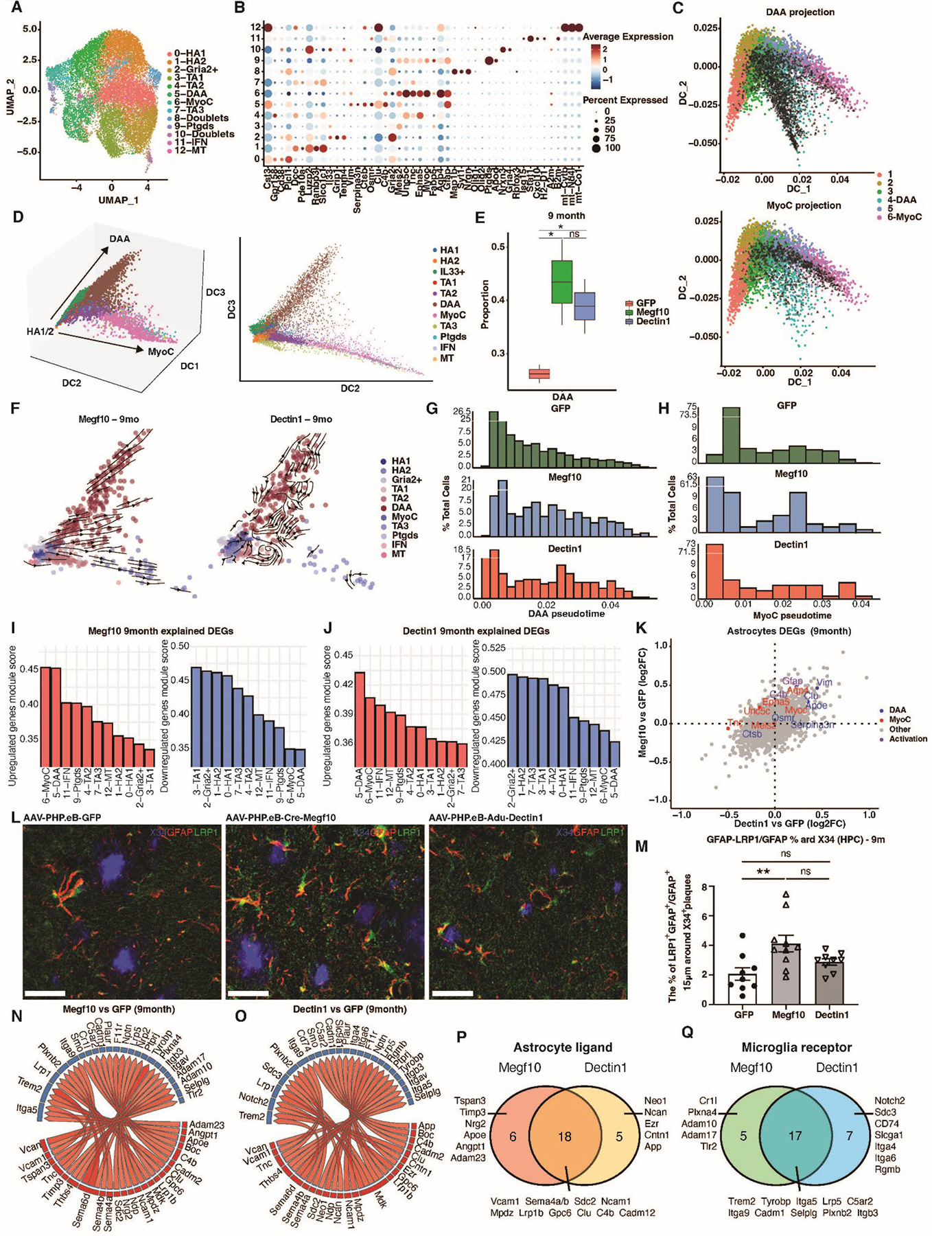
图4. 星形胶质细胞亚群变化(A and B) Diversity of hippocampal astrocytes resolved by snRNA-seq. (A) UMAP of hippocampal astrocyte subsets after subclustering from the total dataset (as in Supplementary Figure 8A; n = 18,492 nuclei). (B) Dot plot of marker genes for astrocyte subclusters. (C) Projection of cluster 5 - DAA (top) and cluster 6 - MyoC astrocyte (bottom) onto diffusion map of snRNA-seq data from Habib et al. Colored dots: nuclei from Habib et al., black dots: nuclei from the present study. (D) 3D (left) and 2D (right) diffusion map embeddings of astrocyte subclusters (as in Figure 5A following exclusion of doublet clusters; total of 17,936 nuclei). Diffusion components 2 and 3 were used for the 2D embedding as they best recapitulate the differentiation trajectories. (E) Proportions of disease-associated astrocytes in GFP, Crene-Megf10, or Adu-Dectin1-treated groups at 9 months old. *: statistically credible change by scCoda. (F) RNA velocity stream plot on 2D diffusion map embeddings of Megf10 and Dectin1 astrocyte subsets at 9 months old (n = 607 and 460 nuclei, respectively). (G and H) Histogram of astrocyte distribution along pseudotime for DAA (G) and MyoC (H) trajectories at 9 months old. (I and J) Bar plot showing module scores of upregulated (left) and downregulated (right) genes in each astrocyte subcluster for Crene-Megf10 vs GFP (I) or Adu-Dectin1 vs GFP (J) at 9 months old. Related to Supplementary Figure 8D–F. (K) Scatter plot of differentially expressed genes in astrocytes for Crene-Megf10 vs GFP and Adu-Dectin1 vs GFP at 9 months old. Representative genes are highlighted in color. (L) Representative images and (M) quantification of the percentage of GFAP+/LRP1+ voxels within GFAP+ astrocytes (%) 15μm around X34+ plaque in the hippocampus of treated 5xFAD mice at 9 months of age. Scale bars: 30 μm. (N and O) Circos plot displaying the top 30 upregulated ligand-receptor interactions in Crene-Megf10 (N) or Adu-Dectin1 (O) compared with GFP at 9 months old. Red: astrocytes; Blue: microglia. (P and Q) Pie graphs of overlapped gene analysis in the ligand-receptor interactions from astrocytic ligands (P) to microglial receptors (Q). HA: homeostatic astrocytes. TA: transitional astrocytes. DAA: disease-associated astrocytes. IFN: interferon-responsive astrocytes. DEGs: differentially expressed genes. Data in M are presented as mean ± SEM. N=9 for GFP, N=10 for Megf10, and N=9 for Dectin1. Statistical significance was calculated using one-way ANOVA with Tukey correction for multiple comparison: *P < 0.05, **P < 0.01, ***P < 0.001, ****P < 0.0001, ns: not significant.(图源:Science. 2026 March 05; 391(6789): eads3972. doi:10.1126/science.ads3972.)
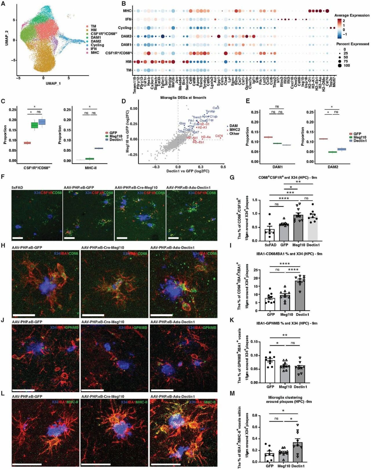
图5. 小胶质细胞应答改变(A and B) Microglia subsets resolved by snRNA-seq. (A) UMAP of microglia subsets following integration of the 5- and 9-month-old timepoints (n = 31,581 nuclei). (B) Dot plot of marker genes for each microglia subset. (C) Box plot showing the proportion of CD68hi (left) and MHC-II (right) microglia subsets at 9 months old. (D) Scatter plot of differentially expressed genes in microglia comparing Crene-Megf10 vs GFP and Adu-Dectin1 vs GFP at 9 months old. Blue: DAM genes. Red: MHC-II genes. (E) Box plot showing the proportions of DAM1 (left) and DAM2 (right) in GFP, Crene-Megf10, and Adu-Dectin1 groups at 9 months old. (F) Representative images and (G) quantification of the percentage of CSF1R+/CD68+ voxels within 15μm around X34+ plaque in the hippocampus of treated 5xFAD mice at 9 months of age. Scale bars: 30 μm. (H) Representative images and (I) quantification of the percentage of CD68+/IBA1+ voxels within IBA1+ microglia (%) 15μm around X34+ plaque in the hippocampus of treated 5xFAD mice at 9 months of age. Scale bars: 30 μm. (J) Representative images and (K) quantification of the percentage of GPNMB+/IBA1+ voxels within IBA1+ microglia (%) 15μm around X34+ plaque in the hippocampus of treated 5xFAD mice at 9 months of age. Scale bars: 30 μm. (L) Representative images and (M) quantification of the percentage of MHC-II+/IBA1+ voxels within IBA1+ microglia (%) 15μm around X34+ plaque in the hippocampus of treated 5xFAD mice at 9 months of age. Scale bars: 30 μm. TM: transitional microglia. HM: homeostatic microglia. DAM: disease-associated microglia. Data in G, I, K, M are presented as mean ± SEM. N=7 for 5xFAD in G, N=9 for GFP, N=10 for Megf10, and N=9 for Dectin1. Statistical significance was calculated using one-way ANOVA with Tukey correction for multiple comparison: *P < 0.05, **P < 0.01, ***P < 0.001, ****P < 0.0001, ns: not significant.(图源:Science. 2026 March 05; 391(6789): eads3972. doi:10.1126/science.ads3972.)
值得注意:尽管病理指标大幅改善,但该研究模型中未观察到明确行为学认知获益,推测可能和 CAR 介导的过度吞噬修剪突触、AAV 载体本身对动物行为的干扰相关,提示后续还需对 CAR 结构、给药剂量进一步优化。
适应症拓展
值得关注的是,CAR-A 这类通过基因工程改造脑内内源细胞、重塑胶质细胞微环境的治疗策略,其疗效评估不能仅依赖 Aβ 斑块负荷的下降,更需要在能同时反映行为学认知与多维度病理改变的模型中进行系统验证,这为适应症拓展与临床转化评价提供了关键支撑。
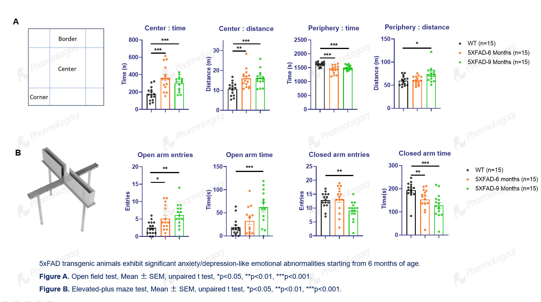
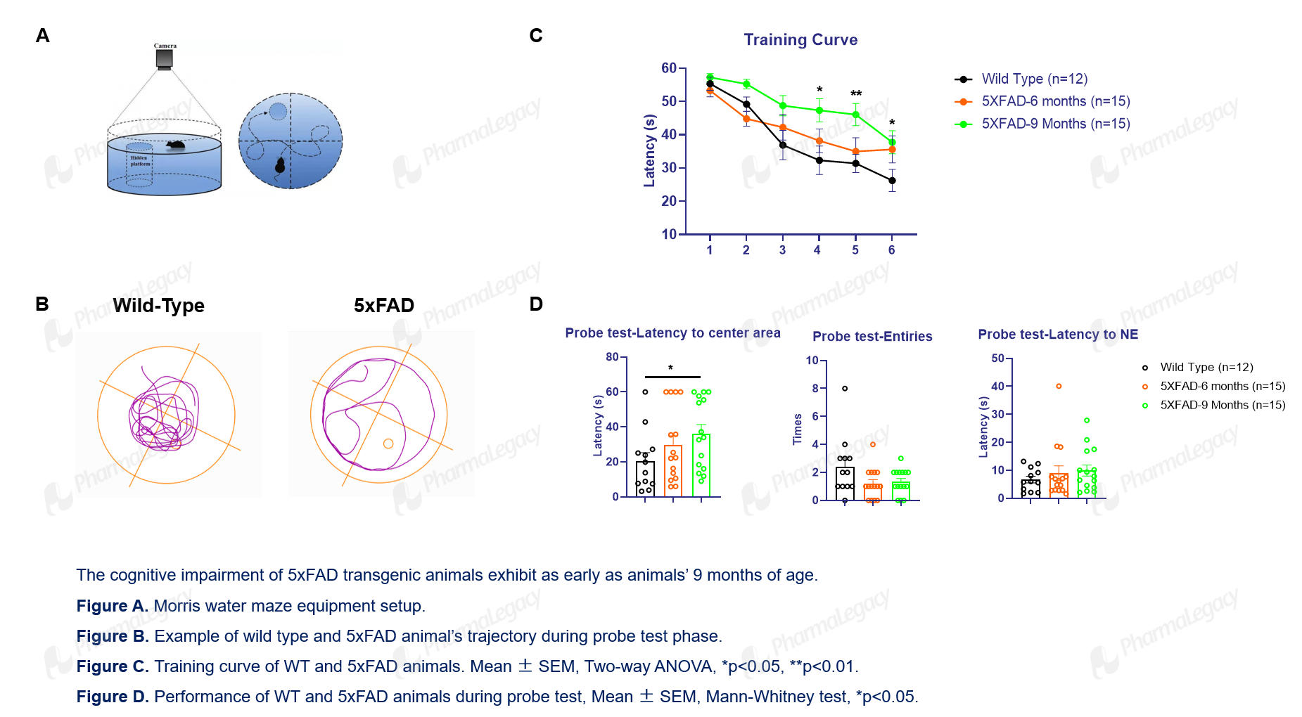
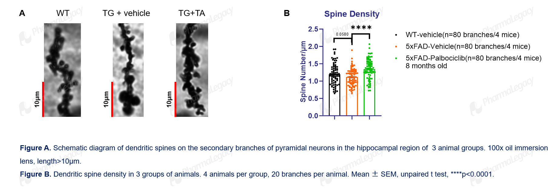
5×FAD 转基因小鼠正是这类新疗法临床前评价的优选工具。作为阿尔茨海默病(AD)研究中应用最广泛的动物模型之一,它过表达携带 3 个家族性 AD 突变的人源淀粉样前体蛋白(APP),同时表达含 2 个致病突变的早老素 1(PSEN1)突变体,可在海马与大脑皮层诱发胞内 Aβ 蓄积、胞外淀粉样斑块沉积、神经炎症、突触损伤及渐进性神经元丢失,并随病程呈现进行性认知衰退,甚至伴有焦虑抑郁样情绪异常、树突棘密度下降等多维度神经病理表型。该品系发病早、病理进展快、实验重复性好,且与人源淀粉样病理具有较高转化参考价值,可同时用于解析 AD 发病机制、评估神经保护与抗淀粉样蛋白治疗策略,适配基因治疗、抗体、小分子等不同类型 AD 候选药物的综合药效评价。
基于这一模型,澎立生物搭建了完善的 AD 临床前药理药效评价体系:通过莫里斯水迷宫、新物体识别实验、Y 迷宫等一系列行为学检测,评价受试疗法对模型动物学习记忆损伤的改善作用;实验终点结合组织病理学、分子生物学与离体电生理开展系统性评估 —— 组织病理染色可对海马、大脑皮层等关键脑区的淀粉样斑块沉积、神经元丢失、突触结构完整性及神经炎症进行定量分析;Western blot、实时荧光定量 PCR 等分子实验可检测 Aβ 相关蛋白、炎症因子与突触标志物的表达变化,阐明药效背后的分子机制;离体脑片电生理用于记录海马长时程增强(LTP)与长时程抑制(LTD),直接反映与认知功能密切相关的突触可塑性改变,为 CAR-A 等中枢神经创新疗法提供从分子到行为的完整证据链。
1.行为——焦虑与抑郁(旷场和高架十字迷宫)
2.行为——莫里斯水迷(MWM)测试
3.高尔基染色——树突棘密度
澎立生物 CNS 平台,助力神经退行性疾病创新疗法临床前评价
针对 AAV 基因治疗、抗体、细胞疗法等中枢创新药物研发痛点,澎立生物搭建完整中枢神经系统(CNS)药效评价平台,覆盖从早期分子筛选到体内药效验证全链条服务。
血脑屏障相关评价:脑脊液、脑组织药物分布检测,评估中枢递送效率,支持AAV、抗体、多肽等多种分子的脑内递送评估。
神经退行性疾病模型体系:拥有5xFAD、APP/PS1等多种阿尔茨海默病模型,同时覆盖帕金森、ALS等多种神经退行性疾病动物模型;支持转基因、病毒、化学诱导造模,适配基因治疗、细胞治疗新型疗法的药效评价需求。
多维药效证据链:行为学功能评估、在体电生理记录、活体分子影像学检测、组织病理学分析、突触标志物检测、小胶质/星形胶质细胞免疫荧光表征、Aβ/tau 生物标志物 ELISA 定量、单细胞转录组机制解析等多维度技术平台;可支持静脉注射、立体定位、侧脑室、延髓池等多种中枢给药途径的临床前药效学评价。
如果您正在开展阿尔茨海默病、其他神经退行性疾病创新药物(抗体、基因治疗、细胞治疗)临床前研发,欢迎联系澎立生物获取定制化的CNS药效评价方案。
原文DOI:10.1126/science.ads3972
免责声明:
本文仅作科学信息与行业交流之用,文中观点及研究结论来源于公开资料,不代表澎立生物立场,亦不构成医疗建议、治疗方案推荐或用药指导。如有疾病诊疗需求,请前往正规医疗机构并咨询专业医务人员。

在线咨询


请添加“业务小助手”企业微信


电话咨询

业务咨询
中国:
+86-130-0328-0039(业务咨询)
+86(21)6176-5100(总机)
Email:info@pharmalegacy.com
海外:
+1-617-803-9415(海外业务咨询)
+86-021-6176-5100*8051(海外业务咨询)
Email:info@pharmalegacy.com
投资者:
+86(21)6176-5100*8002
Email:IR@pharmalegacy.com

TOP


 返回



 返回列表